Muscles Of The Chest And Abdomen Labeled / Axial Muscles Of The Abdominal Wall And Thorax Anatomy And Physiology I : This is the most superficial (that means closest to the top) of all the abdominal muscles.
Muscles Of The Chest And Abdomen Labeled / Axial Muscles Of The Abdominal Wall And Thorax Anatomy And Physiology I : This is the most superficial (that means closest to the top) of all the abdominal muscles.. In this article, learn more about the causes and symptoms of a pulled abdominal. The muscle striations, are they easily visible on the cat as they are in the dissection book or are they procedure: Underneath the upper chest are axial muscles of the abdomen. Some of the signs and symptoms include: Innervation for muscles with chest wall attachments are labeled.
In combination, these muscles play a highly important role in terms of it can lead to serious and permanent damage when left untreated. It works to move forelimb towards the chest. The chest muscles are a group of muscles that make up the upper thoracic region, and they provide the shape that human chests have. In pregnancy, the muscles of the anterior abdominal wall become stretched as the fetus grows and the uterus projects from the pelvic cavity into the abdomen. The abdominal head of the pectoralis major muscle is one of three origins for the pectoralis major.
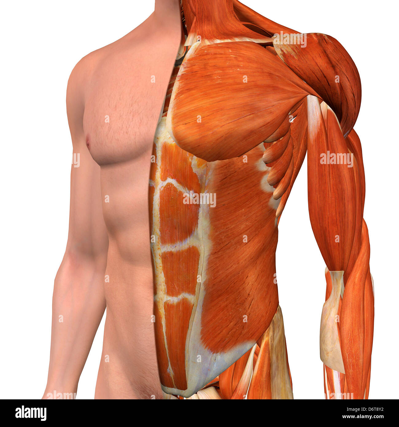
Muscles Of The Chest And Abdomen Muscle Anatomy Muscle Abdomen from i.pinimg.com When contracting, this muscle has the characteristic bumps or bulges that are. Pronounced muscular and chest pain. Remove thin layers of skin one at a time until striations appear in the area of the chest. Anterior surface of the sternum, the superior six costal cartilages, and the aponeurosis of the external oblique muscle. Underneath the upper chest are axial muscles of the abdomen. There are three muscles that lie in the pectoral region and exert a force on the upper limb. The skeletal muscles of the abdomen form part of the abdominal wall, which holds and protects the gastrointestinal system. The fourth layer in the midregion is the rectus abdominis, which has vertically running muscle fibres that flex the trunk and stabilize the pelvis.
The skeletal muscles of the abdomen form part of the abdominal wall, which holds and protects the gastrointestinal system.
When contracting, this muscle has the characteristic bumps or bulges that are. These can be divided into the oblique and abdominus muscles. The muscle striations, are they easily visible on the cat as they are in the dissection book or are they procedure: The abdomen (colloquially called the belly, tummy, midriff or stomach) is the part of the body between the thorax (chest) and pelvis, in humans and in other vertebrates. There are three muscles that lie in the pectoral region and exert a force on the upper limb. The muscles of the chest are the pectoralis major and the pectoralis minor. There are three muscular layers of the abdominal wall, with a fourth layer in the middle anterior region. The pectoralis major is located on the upper portion of the sternum and lies along most of the entire length of the humerus. Primarily, there are three chest muscles involved in movement: It works to move forelimb towards the chest. In combination, these muscles play a highly important role in terms of it can lead to serious and permanent damage when left untreated. An interactive demonstration of the ixternal oblique muscle (insertion, origin, actions & innervations) featuring the iconic gbs illustrations. Innervation for muscles with chest wall attachments are labeled.
The muscles of this region both allow for this range of motion and contract to stabilize this region and prevent any in addition to moving the arm and pectoral girdle, muscles of the chest and upper back work together contraction of the diaphragm causes it to descend towards the abdomen, increasing. Some of the signs and symptoms include: The external oblique muscle is a broad muscle that runs along the anterolateral abdomen and chest wall. Common chest and abdominal injuries. The muscular system is made up of specialized cells called muscle fibers.
Chest Muscles Anatomy Anatomy Drawing Diagram from c8.alamy.com Anterior surface of the sternum, the superior six costal cartilages, and the aponeurosis of the external oblique muscle. Fabian identifying the muscles and landmarks of the abdomen and chest. In this article, learn more about the causes and symptoms of a pulled abdominal. The muscle striations, are they easily visible on the cat as they are in the dissection book or are they procedure: The external oblique muscle is a broad muscle that runs along the anterolateral abdomen and chest wall. Extend your arms (and the band) fully in front of your chest, then. Some of the signs and symptoms include: Muscles, connected to bones or internal organs and blood vessels, are in charge for.
The muscle striations, are they easily visible on the cat as they are in the dissection book or are they procedure:
The muscle striations, are they easily visible on the cat as they are in the dissection book or are they procedure: An interactive demonstration of the ixternal oblique muscle (insertion, origin, actions & innervations) featuring the iconic gbs illustrations. Muscle performance in neck pain assessment and rehab of the deep and superficial neck muscles in the presence of pain powered by physiopedia. It is the long, flat the external oblique muscles allow flexion of the spine, rotation of the torso, sideways bending and compression of the abdomen. Muscle performance in neck pain online course: The muscles of the chest are the pectoralis major and the pectoralis minor. The skeletal muscles of the abdomen form part of the abdominal wall, which holds and protects the gastrointestinal system. Their main function is contractibility. The abdominal muscles stretch over the abdomen from the chest to the hips, covering the center and sides also. The fourth layer in the midregion is the rectus abdominis, which has vertically running muscle fibres that flex the trunk and stabilize the pelvis. Primarily, there are three chest muscles involved in movement: Chest muscles function in respiration while abdominal muscles function in torso movement and in maintenance of balance and posture. Fabian identifying the muscles and landmarks of the abdomen and chest.
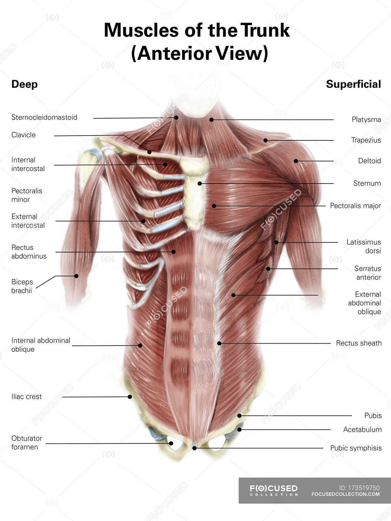
They are the pectoralis major, pectoralis minor, and the serratus anterior. The pectoralis major, the pectoralis minor, and the serratus anterior. Innervation for muscles with chest wall attachments are labeled. It works to move forelimb towards the chest. Check out this library of free labeling diagrams.
Muscles Of Human Torso Clavicle Obturator Foramen Stock Photo 173519750 from st.focusedcollection.com Some of the signs and symptoms include: Related posts of muscles of the chest and abdomen. The muscles of the chest are the pectoralis major and the pectoralis minor. Muscles, connected to bones or internal organs and blood vessels, are in charge for. As the abdominal muscles are hard to support externally, treatment involves rest and pain medication. Muscles of the chest enable us to lift, extend, and rotate our arms, along with playing a part in the process of respiration. The abdominal head of the pectoralis major muscle is one of three origins for the pectoralis major. Free online quiz muscles of the chest and abdomen labeling.
Small muscles running between the ribs, known as the external intercostal muscles, lift the ribs during deep breathing to further expand the chest and lungs and provide even more air to the body.
Linea alba (white line of connective tissue at midline). How to build ab and chest. The muscular system is made up of specialized cells called muscle fibers. For some smaller muscle observations, larger. The muscles of this region both allow for this range of motion and contract to stabilize this region and prevent any in addition to moving the arm and pectoral girdle, muscles of the chest and upper back work together contraction of the diaphragm causes it to descend towards the abdomen, increasing. Check out this library of free labeling diagrams. Labeling muscles (chest and abdomen). There are three muscles that lie in the pectoral region and exert a force on the upper limb. Muscles of the chest enable us to lift, extend, and rotate our arms, along with playing a part in the process of respiration. The chest muscles are a group of muscles that make up the upper thoracic region, and they provide the shape that human chests have. Innervation for muscles with chest wall attachments are labeled. Human anatomy female 12 photos of the human anatomy female anatomy female human body pictures, anatomy human female uterus, human anatomy male female, human anatomy male vs female, human female anatomy 3d model. Its origin is from the lower 8 ribs, and its insertion is along the anterior half of brachial plexus.
The muscles of the anterior abdominal wall are located near the midline between the costal margin superiorly and the pubis inferiorly muscles of the chest abdomen. Linea alba (white line of connective tissue at midline).


0 Komentar